Have you ever asked a neighbor for something? Maybe you needed a cup of milk or a few eggs. Perhaps you inquired about borrowing a lawn tool. You may have asked for assistance lifting a new piece of furniture into your home.

Image credit: Getty Images
Whatever it was, a neighbor asking for help with some amazing DNA science probably isn’t something you’ve had happen. Interestingly, that’s exactly what occurred at the Science Museum recently and we jumped at the chance to help!
Last month, Dr. Sean Koebley, a postdoctoral fellow with the Virginia Commonwealth University Department of Physics, contacted us about using some specialized equipment in The Forge. The Forge is our makerspace comprised of a workroom and machine shop equipped with numerous high- and low-tech tools, such as vices, a CNC router, drill presses, a 3D printer, a 36” plotter, a belt sander, sewing machines, and more.
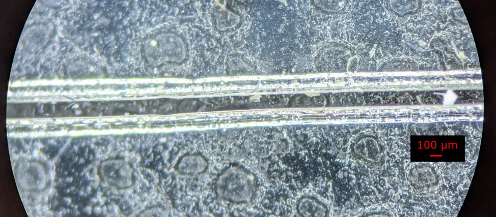
Koebley reached out to Science Museum Maker Education Manager Matt Baker about using The Forge’s precision laser cutter. Koebley needed to cut tiny channels through a film of PDMS (a type of plastic polymer) and acetate on a glass microscope slide. These channels required precision equipment as they were only 100 microns, or 0.1 mm. That’s about the thickness of a sheet of paper!
After removing the acetate layer, which protects the PDMS and glass from carbon and other “junk” left over from the cutting process, Dr. Koebley and his research team created a 1 cm-tall mold of the channels.
Here’s a picture of what these channels look like at micrometer resolution:

That part is really interesting, but what’s more interesting is what the team is doing with the precision-cut items. The researchers will flow DNA strands—yes, that DNA, the coded hereditary material in almost all organisms, including humans—into the molded channels. Doing this forces the strands to elongate instead of spiraling into its regular double-helix structure. By using a high-speed atomic-force microscope, the researchers can get a better look at—and take better images of—longer, uncoiled strands of DNA. These images help them study molecular “barcodes” on DNA.
The human genome is full of repeating sequences. Scientists use a recently developed, programmable enzyme known as CRISPR/Cas9 to attach to repeated sequences in DNA, then take a very high-resolution image the DNA and patterns of attached CRISPR enzymes (the barcode mentioned earlier). This is done to determine what the normal barcode looks like so that when it is compared to a unique DNA sample, scientists can detect genetic differences.

Image Credit: Getty Images
This research is important because it allows scientists to identify genetic changes and mutations more quickly and cheaply, which can facilitate better, earlier treatments of genetic conditions such as sickle cell anemia, cystic fibrosis, and cancer.
For more on this process, here’s the paper (appropriately titled "Rapid fabrication of microfluidic PDMS devices from reusable PDMS molds using laser ablation") on which Koebley based his laser-cut PDMS/acetate slides. VCU has also shared information about the research its scientists are conducting here and here.
Not only is this a perfect real-world display of STEM, but it’s also a chance for the Science Museum to help out its neighbor, VCU. Collaboration breeds innovation, or in this case, the advancement of DNA science!


